SCSCR is primarily focusing on several areas of research in stem cell biology at both cellular and molecular levels such as aging of stem cells, microenvironment–mediated regulation of hematopoietic stem cells, development of 3D cultures using biomaterials, mesenchymal stem cell biology with special focus on exosomes and micro-vesicles and signal transduction, endometrial stem cells, tissue-specific differentiation of human ES cells, etc.

1. Experimental Hematology

I) Microenvironment-mediated regulation of Hematopoietic Stem Cells:

(A) Priming of MSCs to boost their hematopoiesis-supportive ability

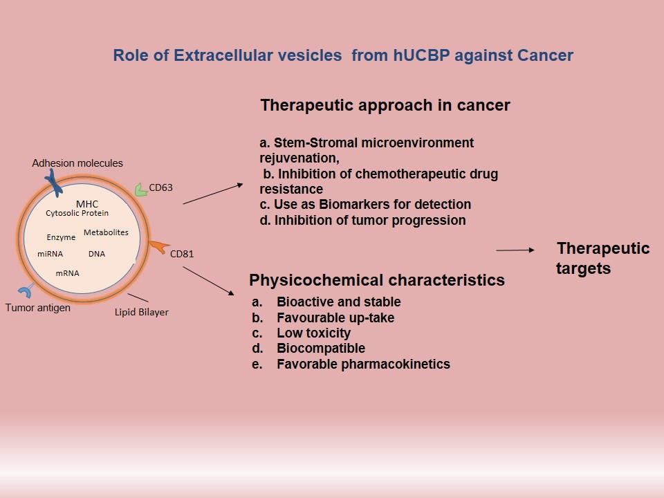
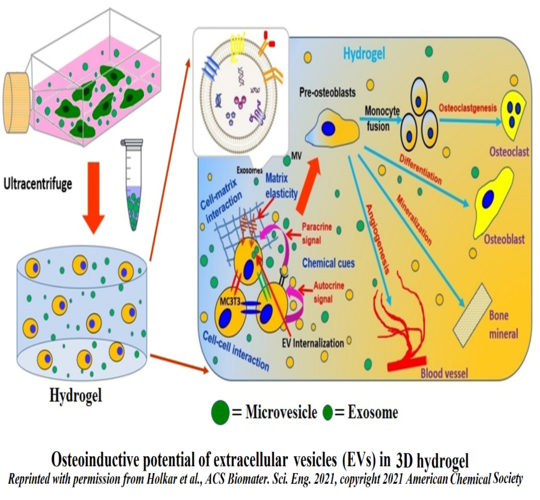
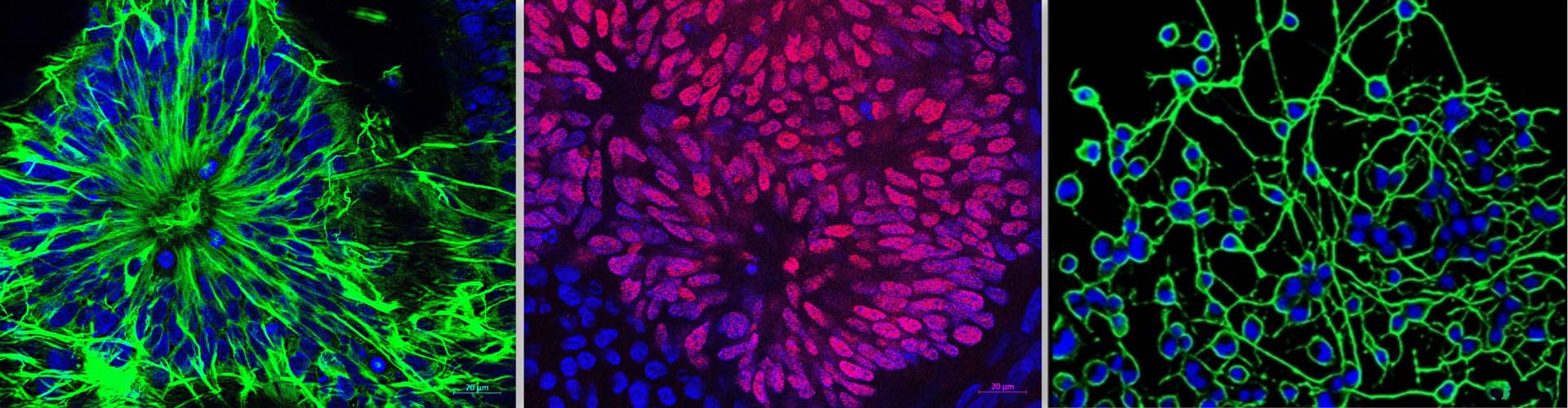
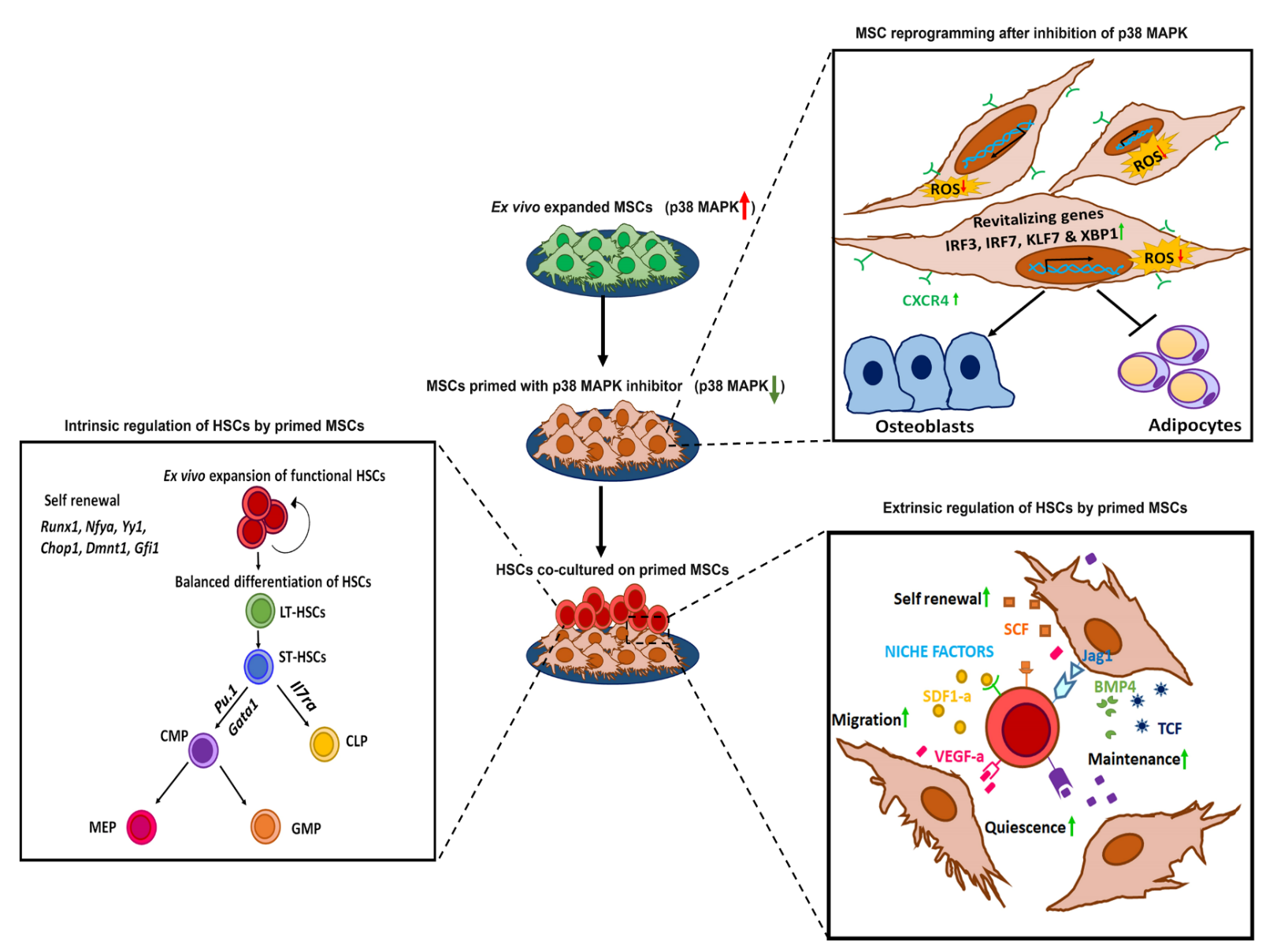
In the bone marrow microenvironment, the mesenchymal stromal cells (MSCs) respond to the external cues and modulate the fate of hematopoietic stem cells (HSCs) via the extracellular vesicles (EVs). The EVs viz. micro-vesicles (MVs) and exosomes, therefore, form an important extrinsic mechanism through which the stromal cells regulate the fate of HSCs.

In this project, we use a strategy known as “priming of MSCs” wherein we modulate the signalling pathways in MSCs, which improves their HSC- supportive ability. Such a strategy could yield a sufficient number of HSCs with superior functionality to overcome challenges associated with hematopoietic stem cell transplantation (HSCT) at the clinical level. Furthermore, priming also rejuvenates the MSCs, thereby improving the overall outcome of in vitro cultured MSCs for regenerative therapies. The EVs isolated from primed MSCs could also be used as off-the-shelf, cell-free biologics in a clinical setup.

Principal Investigator: Dr. Anuradha Vaidya and Dr. Vaijayanti Kale

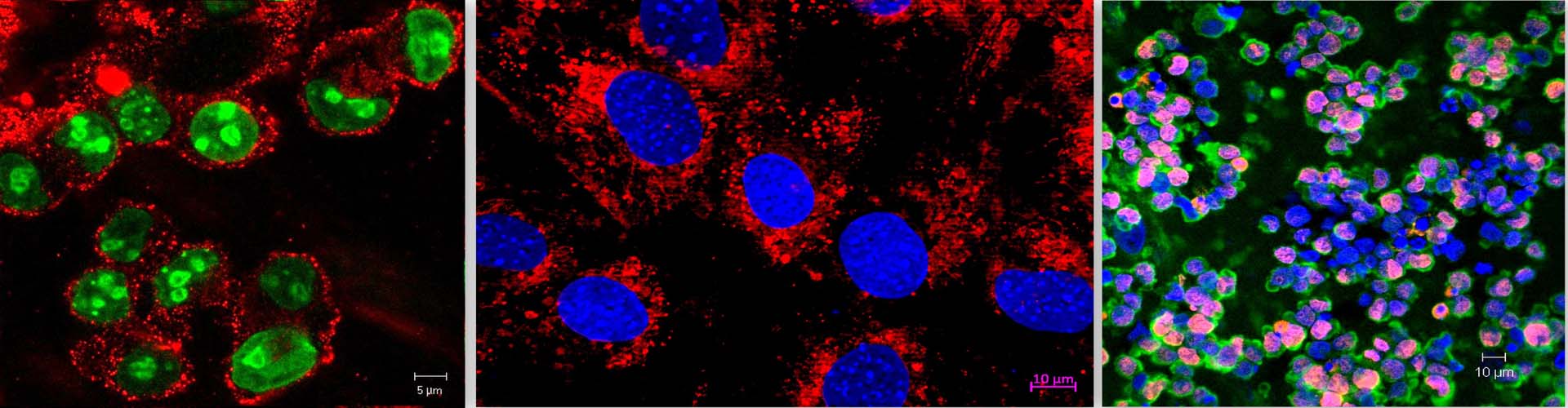
(B) Elucidation of signalling mechanisms induced by EVs secreted by Acute myeloid leukemia blasts having specific mutations

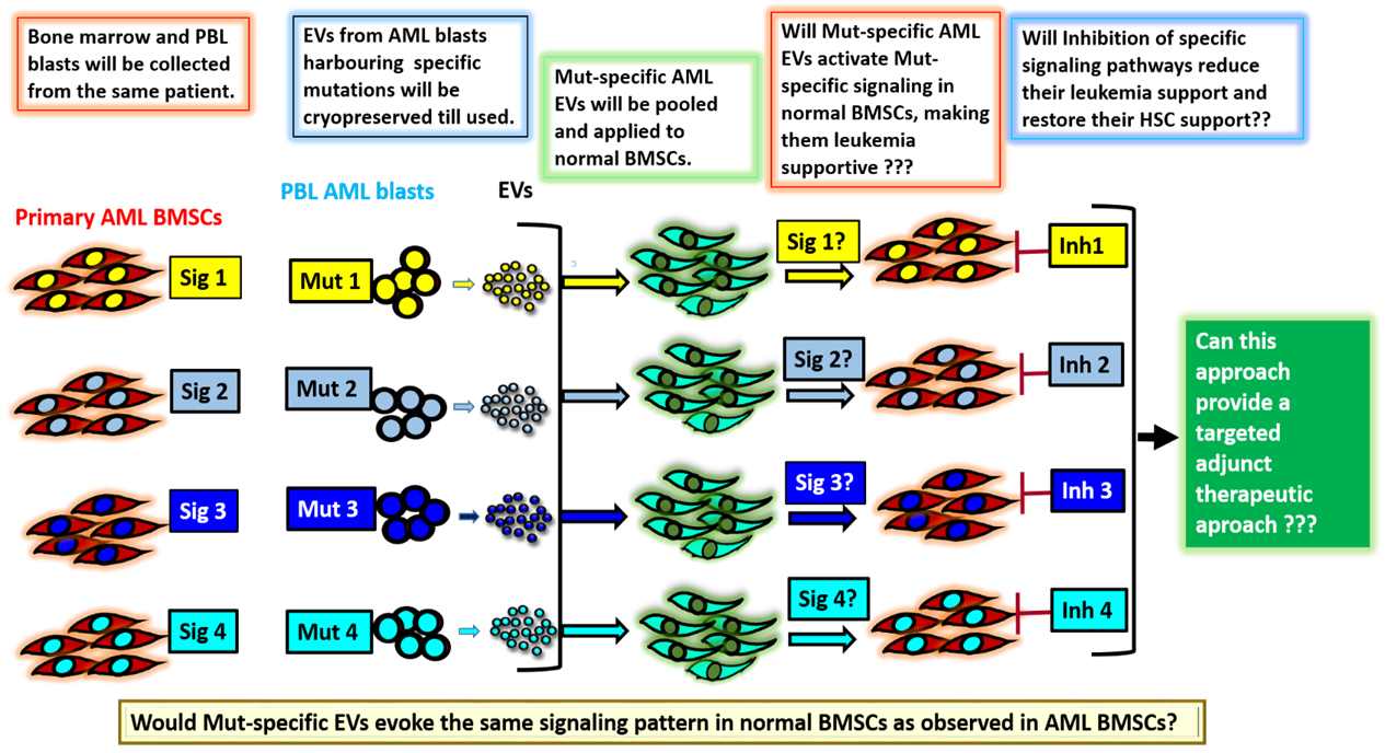
Acute myeloid leukemia (AML) is a type of blood cancer involving specific mutations that can correlate with poor prognosis in patients. In recent years, several studies have underscored the role of the bone marrow microenvironment, especially the MSCs, in the development and progression of the disease. Most of these studies have investigated the effect of the leukemic cells on the bone marrow microenvironment. However, the reverse that is how the leukemic bone marrow stromal cells (BMSCs) modulate the normal HSCs is still not clearly understood. Furthermore, so far, no study has established a correlation between the mutations in the blast cells with the signaling pathways prevailing in the BMSCs. In addition, whether such altered signaling is responsible for conferring a chemo-resistant phenotype on the leukemic stem cells (LSCs), and whether the altered BMSCs lose their HSC- supportive properties has not been investigated. This project focuses on understanding the interplay between the BMSCs and the blasts from AML and aims to decipher how the AML blasts or their extracellular vesicles (EVs) modulate the signaling mechanisms in the marrow stromal cells to their advantage. Such an understanding is crucial because one of the hallmarks of AML is over-production of the immature blasts, which crowd the bone marrow, preventing it from making normal blood cells. Such studies could provide an impetus in designing novel targeted therapies against AML that could be less toxic and more specific.

Principal Investigator: Dr. Anuradha Vaidya and Dr. Vaijayanti Kale

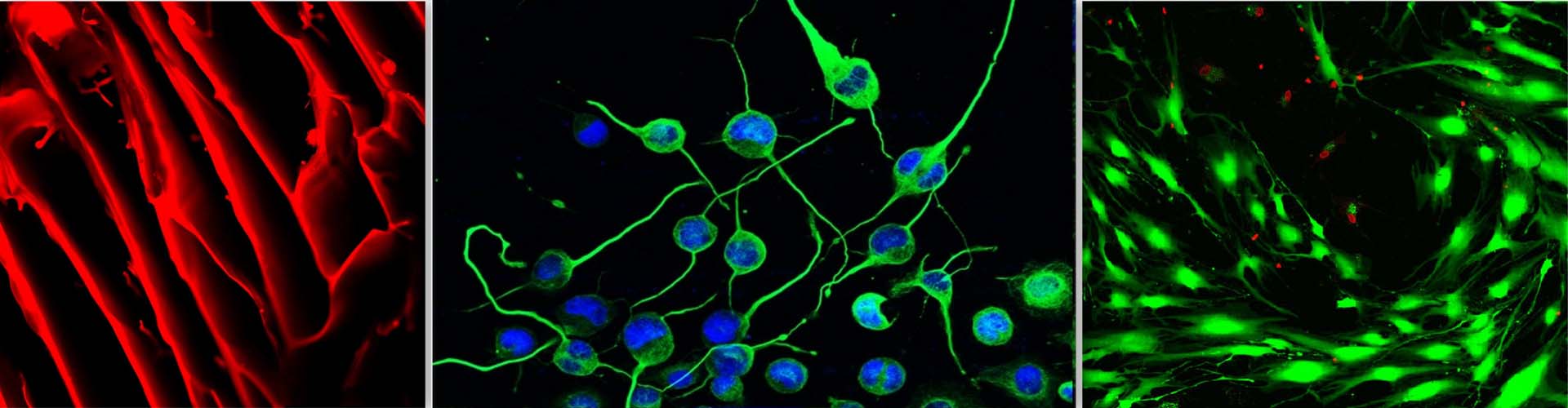
II] Neuroprotective effect of secretome derived from bone marrow-derived MSCs

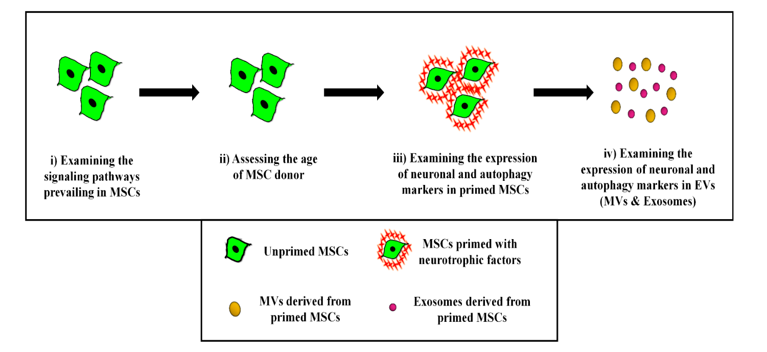
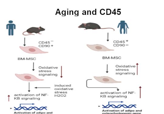
Neurodegenerative diseases (ND) are characterized by a progressive and irreversible loss of neuronal cells leading to cognitive impairments and memory loss. Difficulties in diagnosis and lack of validated in vitro and in vivo models impose challenges in drug discovery and development against ND. Mesenchymal stromal cells (MSCs) are being explored as cell therapy for various ND, but because of their large size they get trapped into primary filter organs like lungs and liver, and also cannot pass the blood-brain barrier (BBB). However, it has been reported that even after being trapped in filter organs, MSCs exert their salutary effects on distant tissues in a paracrine manner through their secretome. Hence, in the last few years, the focus of research has slowly shifted from MSC-based cellular therapy towards harnessing the regenerative potential of the MSCs’ secretome. The primary aim of this project is the establishment of validated oxidative stress and endoplasmic stress-induced-neurodegenerative model systems to examine the salutary effect of the MSC secretome. We would also like to examine whether in vitro priming of young MSCs with neurotrophic factors or certain pharmacological reagents like nitric oxide donors would boost their potential to reverse the loss of neurogenesis induced by stress or aging.

Principal Investigator: Dr. Anuradha Vaidya and Dr. Vaijayanti Kale

2. Aging

Rejuvenating effects of Extracellular vesicles on aging

There is a growing number of aged individuals in the global population. Aging and the chronic diseases associated with it place a tremendous burden on our healthcare system. Recently, Extracellular vesicles from different sources are considered as a new weapon to fight against aging-mediated hematological and non-hematological disorders.

For the aged people it is often challenging to retain a high level of physical and mental capacity. During aging, many elderly patients suffer from several aging-mediated hematological and non-haematological disorders. Mesenchymal stem/stromal cells (MSCs) hold promise in regenerative therapies due to their multi-potency and easy availability. High prevalence of musculoskeletal diseases found in elderly population has motivated researchers to elucidate the impact of aging on MSC properties, as the age of the donors appears to be one of the most important parameters that affects the MSC properties. It is therefore, very important to elucidate the fundamental mechanisms of aging for development of strategies to minimize the impact of aging on our health and economy.

Recently, Extracellular vesicles (EVs) have emerged as important mediators of intercellular communication, playing a critical role in modulating hematopoiesis within the bone marrow microenvironment. It has been demonstrated that human umbilical cord blood-derived plasma (hUCBP) has unique anti-aging effects. Here in this current project, we would examine whether EVs from the hUCBP could play an important role in modulation of age-related symptoms.

Principal Investigator: Dr. Madhurima Das

Co-Investigators: Dr. Vaijayanti Kale, Dr. Anuradha Vaidya and Dr. Jyotsna Potdar

3. Cancer Biology

Rejuvenating effects of cord blood- / young MSC-derived extra-cellular vesicles on chemotherapy-induced damage of bone marrow mesenchymal stromal cells: in vitro and in vivo studies using an experimental AML mouse model.

To all of us, cancer is perhaps the most frightening of diseases. Acute myelogenous leukemia (AML) is a heterogeneous disease characterized by a blockade in the differentiation of hematopoietic stem cells and a clonal expansion of myeloid blasts in the bone marrow and peripheral blood. Despite several therapeutic approaches like radiotherapy, chemotherapy, etc., many patients show resistance to such therapeutic measures. In most cases, AML patients undergo relapse and succumb to the disease after chemotherapeutic measures.

Studies showed that both murine and human MSC-derived EVs were able to ameliorate radiation-induced damage to murine bone marrow cells by blocking the radiation-induced growth inhibition, DNA damage, and apoptosis.

Here in this present study, we propose to determine whether cord blood plasma (CBP)-derived or young BMSC-derived EVs can reverse the chemotherapy-induced bone marrow stem/stromal cell damage. EV-mediated reversal of long-term damage of marrow stem cells after chemo-induction may form a unique new therapeutic approach against chemo-induced damage.

Principal Investigator: Dr. Madhurima Das

Co-Investigators: Dr. Vaijayanti Kale, Dr. Anuradha Vaidya and Dr. Jyotsna Potdar

4. Biomaterials and Stem Cells in Regenerative Medicines

The design of biomaterial and the supply of physical, as well as biological stimuli, play an important role in regulating cell function to create a tissue implant for clinical applications. Developing bone and cartilage biomaterials with material compositions, geometric structures, and physiological functions such as natural bone and investigating their interactions with stem cells for bone repair remain significant avenues for future research. At SCSCR we use various natural and synthetic materials, mechanical/or biological stimulation, and cutting-edge synthesis techniques to develop an engineered construct that meets the functional and biological demands of native tissue. The SCSCR team aims to discover new strategies for accelerating the repair and regeneration of lost, damaged, or diseased tissues (especially bone and cartilage) and translate these findings to help animal and human patients in need.

The goal of SCSCR is to design polymeric biomaterials for specific orthopaedic applications. At SCSCR we focus on developing synthetic and naturally-derived hydrogel biomaterials in conjunction with biochemical and mechanical stimuli to promote priming of stem cells to express a particular phenotype, as well as deliver stem cell–derived extracellular vesicles to promote healing of damaged or diseased tissues. In terms of our tissue engineering and regenerative medicine efforts, at SCSCR we primarily focus on bone and cartilage regeneration.

Biomimetic and bilayered interpenetrating network (IPN) hydrogel for osteochondral tissue regeneration

This project focusses on developing a novel class of interpenetrating network (IPN) hydrogels with a unique combination of mechanical strength and biocompatibility for bone and cartilage tissue regeneration applications. The SCSCR team is working on combining natural (agarose, hyaluronic acid, chondroitin sulphate) and synthetic polymers (PEGDA) along with chondrocytes and osteoblasts in their respective layers based on IPN hydrogels, with a unique blend of properties complementary to nucleated cell growth. The novel signalling approach will be the introduction of hyaluronic acid (HA), RGD, transforming growth factor beta (TGF-β1), and chondroitin sulphate as, cartilage molecules known for their ability to promote chondrogenesis and bone morphogenetic protein-2 (BMP-2) to promote osteogenesis along with hydroxyapatite (HAp) coated microspheres to provide nucleation sites for secreted calcium and accelerated bone formation. These bi-layered IPNs with significantly improved mechanical and biological properties could be used to provide a chondrogenic (for cartilage) and osteogenic (for bone) signalling approach. The outcomes of this proposed work will provide a rationale for in vivo experiments, which is, of course, the necessary route to move on to translational research to bring this technology to researchers, clinicians, practitioners e.g. orthopaedic surgeons, involved in treating arthritis-related diseases.

Principal Investigator: Dr. Ganesh Ingavle

Synthesis of novel polysaccharide-based micro-porous polyHIPE scaffolds for osteochondral tissue regeneration.

The overall objective of this proposal is to develop highly porous scaffolding biomaterials by High Internal Phase Emulsion (HIPE) templating polymerization and to understand how synthesis parameters and incorporation of bioactive signals improve material properties concerning improving cellular response for osteochondral (bone-cartilage) tissue regeneration. At SCSCR we are developing novel 3D polyHIPE scaffolds that possess advantages over current scaffold preparation methods, using water at low temperatures to generate an interconnected pore structure. The SCSCR lab evaluates the in vitro efficacy of these innovative polyHIPE scaffolds for osteochondral (bone-cartilage) tissue regeneration using MC 3T3 (an osteoblast precursor cell line) and chondrocytes obtained from Wistar rat femur condyles. The overall significance of this work will be in introducing and developing a new strategy and biomaterials for musculoskeletal tissue engineering.

Principal Investigator: Dr. Ganesh Ingavle

Constructing cell-free osteoinductive and osteoconductive biomaterials for bone tissue engineering

This project is focusing on constructing cell-free osteoinductive and osteoconductive biomaterial for bone tissue engineering with enhanced mechanical strength and mineralized microenvironment. We are working on developing a 3D-in vitro hydrogel model to assess the functionality of respective components of stem cell secretomes, extracellular vesicles (EVs), by encapsulating them along with MC3T3 cells (a pre-osteoblast cell line) into mineralized- interpenetrating networks IPN hydrogel and thereby studying osteogenic differentiation of encapsulated cells. The novelty of this proposal is the increased mechanical strength of IPN scaffolds as well as a cell-free approach that can result in a commercial 3D osteo-inductive scaffold. The findings of this study will provide a rationale for in vivo studies, which are of course, the required avenue for translational research to introduce this cell-free approach /technology to clinicians and orthopaedic surgeons for an immediate solution for bone regeneration.

Principal Investigator: Dr. Ganesh Ingavle

Co-Investigator: Dr. Vaijayanti Kale

Extracellular vesicles loaded injectable hydrogels

Injectable systems for stem cell transplantation have the advantage of allowing minimally invasive surgical procedures to be used, and thus causing less pain to the patients. The recent trend indicates that there is a shift from the direct usage of Mesenchymal Stem Cells (MSCs), towards the application of the paracrine factors and extracellular vesicles (EVs) isolated from their MSC secretome in bone tissue regeneration. This shift has directed research into the development of “cell-free” therapeutics, which could be a better alternative due to its several advantages over the use of their parental MSCs. At SCSCR we are developing a novel composite injectable hydrogel scaffold system based on RGD peptide modified alginate and hyaluronic acid (HA) to examine its mechanical, biological and osteoconductive performance for applications in bone tissue engineering. Further, the SCSCR team is aiming to replace the cellular components by extracellular vesicles to develop “cell-free therapy”. The osteo-inductive capacity of this cell-free injectable hydrogel system to promote bone formation will be assessed in vitro and in vivo. Outcome from this work will facilitate the introduction of a novel approach using composite injectable hydrogel containing EVs to create native tissue mimicking biomaterials to bone research and other areas of tissue engineering where a slow cell turnover and multiple tissue controlling approach is required to optimize biomimicry. The application of mechanically strong, biomimetic and injectable hydrogel scaffold with improved bioactivity, with an ease incorporation of bioactive signals, growth factors and EVs to accelerated tissue regeneration has a wide range of potential application and will be of interest to a wide range of academic researchers and companies.

Principal Investigator: Dr. Ganesh Ingavle

Co-Investigator: Dr. Vaijayanti Kale, Dr. Prasad Pethe

5. Role of Mechanobiology in Human Embryonic Stem Cell Differentiation

We are uncovering the role of mechanosensors YAP/TAZ during differentiation of human embryonic stem cells into endoderm on substrates of varied stiffness.

Adherent cells such as embryonic stem cells have mechanisms for detecting the substrate stiffness along with numerous cytokine receptors in order to proliferate or differentiate. YAP and TAZ are transcriptional regulators (part of Hippo pathway) that control genes responsible for cell proliferation, migration and cell death in response to the cell substrate stiffness. Substrate stiffness alone has been shown to regulate differentiation of mesenchymal stem cells, with stiffest substrate leads to osteogenic differentiation while lower stiffness leads to adipogenic differentiation. Stiffer substrates activate YAP/TAZ activity and has been shown to play a crucial role in maintaining undifferentiated state of human pluripotent stem cells (hPSCs). Soft substrate has been shown to induce differentiation of hPSCs into neuronal lineage. However, whether YAP/TAZ plays a role in endoderm differentiation of human embryonic stem cells is yet to be demonstrated. Currently, we are investigating the role of YAP/TAZ during endoderm differentiation of human embryonic stem cells in response to varied substrate stiffness.

Principal Investigator: Dr. Prasad Pethe

6. Human Pluripotent Stem Cell Differentiation & Epigenetics

Polycomb group (PcG) proteins along with other epigenetic modulators catalyse histone modifications at promoters and thus they control the expression of genes essential for lineage specification.

Human pluripotent stem cells are the cells with miraculous ability to self-renew and differentiate into all three-germ lineages and have immense potential in the fields of regenerative medicine or cell-based therapies to treat several human disease conditions. However, decades of research has shown that transforming pluripotent stem cells into a mature functional tissue is a difficult task. Thus it in order to decipher this intricate process, we have to understand the mechanism of cell differentiation and maturation. Currently we are pursuing research to understand how Polycomb group (PcG) proteins respond to differentiation cues and add inhibitory histone modifications (H3K27me3& H2AK119ub1) are promoters of crucial genes in human pluripotent stem cells. We are also investigating the role of histone deubiquitinases (H2A- DUBs) that remove the histone mark during human pluripotent stem cell differentiation into neuronal lineage.

Principal Investigator: Dr. Prasad Pethe

Co-Investigators: Dr. Vaijayanti Kale

7. Epigenetics of Endometriosis

Epithelial to mesenchymal (EMT) transition enables endometrial tissue to move to ectopic sites. We hypothesize that Polycomb group (PcG) proteins may mis-regulate the EMT genes leading to endometriosis.

Endometriosis is a condition in which functional endometrial glands and stroma grow outside of the uterine cavity and this growth leads to complications such as dysmenorrhea, dyspareunia, chronic pelvic pain, adhesions and infertility, with fibrosis routinely observed and this further contributes to pain and infertility. However, the epigenetic mechanism that leads to EMT and fibrosis in endometriosis is poorly understood. Polycomb Group (PcG) proteins are histone modifiers that can control the expression of genes involved in EMT by catalysing histone modifications; their misexpression is reported in several cancers. We hypothesise that misregulation of PcGs could lead to overexpression of EMT related genes, which may help endometrial cells migrate and cause fibrosis ectopically. To determine this, we plan to study if the PcGs protein occupy promoters and regulate EMT and fibrosis genes in ectopic endometrial cells as well when specific PcG (RING1B and EZH2) genes are repressed.

Principal Investigator: Dr Prasad Pethe

Co-Investigators: Dr. Vaijayanti Kale, Dr. Anuradha Vaidya, Dr Prasad Bhanap and Dr Sneha Deokar

8. WNT Signalling in Hypertrophic Scar Formation in Post Burn Injuries

Thermal injuries can lead to hypertrophic scars, which currently do not have effective treatment. Using patient scar tissue, we are investigating whether activation of inhibition of WNT signalling can regulate ECM protein production by scar dermal fibroblasts.

Skin tissue plays multiple essential roles such as protection against invasion of microorganisms, prevention of water loss, thermoregulation and mechanosensation; and any injury to the skin tissue can disrupt these functions. Skin is a composite organ with functional co-ordination between various cell types. Trauma such as thermal burns disrupts the skin architecture and in response to the injury, cells such as fibroblasts, macrophages, platelets, and keratinocytes are mobilized to seal the damage, resulting in a scar formation. However, in many cases of severe thermal injury the scar tissue persists. This scar tissue cannot carry out thermoregulation, mechanosensation efficiently and it also restricts motion; currently, there are only few ways to reduce scarring such as pressure garments, surgery and or steroid treatment.

Hence, it is essential that the molecular mechanism driving abnormal scar tissue be studied, so that we can identify molecular targets to reduce abnormal scar tissue formation. Signalling pathways such as – TGF-β, NOTCH, WNT, SONIC HEDGEHOG, HIPPO are involved in embryonic skin development and in adult skin maintenance. WNT signalling pathway is developmentally conserved signalling pathway that plays a crucial role in embryonic development as well as regulation of adult stem cells to maintain tissue homeostasis. Prolonged activation of WNT signalling negatively influences cutaneous wound healing in animal models, however, such data on role of WNT pathway in abnormal scar tissue in humans post thermal injury is unavailable. This project is aimed to understand the effect of WNT signalling pathway activation and inhibition on ECM protein expression in fibroblasts derived from hypertrophic scar tissue that form after thermal injury.

Principal Investigator: Dr. Prasad Pethe

Co-Investigators: Dr. Mugdha Pradhan

9. Infertility

Prior to embryo implantation into the endometrium, there is communication via Extra Cellular vesicles (EVs). Break down in this communication may lead to failure to implantation and subsequently infertility. We are assessing the effect of placental EVs on the signalosome in endometrial cells derived infertile women.

The fetal trophoblasts cells secrete EVs than help embryo implantation and later the syncytiotrophoblasts regulate the maternal immune response and help complete pregnancy to term. Moreover, in the cases of infertility, the lack of communication between the fetal and maternal cells could be the reason for preterm delivery or spontaneous abortion or recurrent miscarriages.

The placental EVs might be interacting with maternal cells and regulating expression of key genes relating to inflammation, matrix metalloproteases, hormones and extracellular matrix. We plan to understand how EVs secreted from placental MSCs regulate certain gene(s) by activation/inhibition of pathways such as AKT, MEK, ERK, p38 and SAPK/JNK in MSCs derived from infertile women.

Principal Investigator: Dr. Prasad Pethe

Co-Investigators: Dr. Vaijayanti Kale, Dr. Madhurima Das, Dr. Anuradha Vaidya and Dr. Jyotsna Potdar